
How Your Eyes Work
If you ask most optometrists why we do what we do for a living, the reply will almost always include a love of science, the desire to help others, and our absolute obsession with the eyes. The eyes are one of the body's most unique, fascinating, and complex organs.
Understanding How Your Eyes Work
The ability to see images takes considerable coordination between multiple parts and processes, and even the slightest abnormality can mean blurred vision or complete visual impairment.
Eye Anatomy: The Basics
To understand how the eyes work, you need a basic understanding of eye anatomy. If you’d like to learn about eye anatomy in detail, visit the American Academy of Ophthalmology’s page on Eye Anatomy. For the purposes of this post, we’ll review the basics:
- The outer eye (and inner eyelid) are covered with a thin membrane called the conjunctiva.
- The cornea is at the front of the eye, underneath the conjunctiva.
- Below the cornea is a fluid-filled layer called the anterior chamber. The fluid inside the anterior chamber is called aqueous humor.
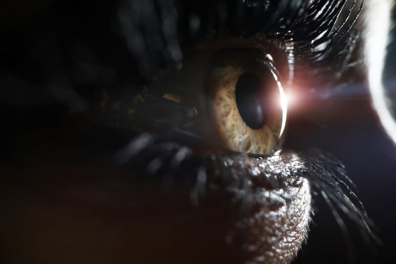
- Underneath those layers are the iris (the colored part of the eye) and the pupil (the black circle in the middle).
- Then there is the lens.
The cornea, iris, pupil, and lens are all at the front portion of the eye. From there, we have the middle of the eye, called the vitreous cavity. The vitreous cavity is filled with a gel-like substance called vitreous humor - which works along with the aqueous humor to maintain proper eye pressure.
Now we make our way into the mid- and rear portions of the eye.
- The retina is a light-sensitive layer of the eye that wraps around the outer portion of the eyeball. The retina is divided into multiple parts. The peripheral retina provides peripheral vision, and the macula provides detailed central vision.
- Rods and cones are specialized cells on the retina that differentiate color.
- The optic nerve connects to the eye at the rear and connects the eyes to the visual cortex area in the brain.
If you’re familiar with some of the most common eye conditions, you may recognize some of these parts by their diagnosis. For example, conjunctivitis - inflammation of the conjunctiva - is one of the most common causes of red, irritated eyes. Macular degeneration is one of the most common causes of age-related vision loss, resulting from a compromised macula. Other common eye issues named for eye anatomy include things like corneal abrasions and retinitis.
Vision Is A Five-Step Process
The anatomical parts of the eye work closely together to bring the world into focus to the best of their ability. If something isn’t working as it should, it’s detected during an eye exam. With accurate diagnosis, we hope that corrective lens prescriptions, medications, or laser surgeries can correct the issue enough to restore vision to a functional state.
Here are the three steps required to create a picture of what you’re looking at in your mind’s eye.
Light enters through the cornea
Light enters through the cornea at the front of the eye through the pupil. The iris acts as a gatekeeper of sorts - opening and closing, which makes the pupil bigger or smaller, regulating the amount of light that passes through. This mechanism protects the eye from absorbing too much light - via UV rays - that damage the eye.
If you look in the mirror in a bright room, you’ll notice the pupil appears very small, like a pinpoint. However, if you close the blinds, dim the light, or move into a darker space, the iris opens - making the pupil larger - to let more available light in.
The cornea and lens bend the light
The light entering the eye is bent by the cornea (refraction), after which the light goes through the aqueous humor. This is the process that brings what you see into focus.
Light is refracted a second time through the lens onto the retina
As the light continues moving through the front of the eye, the lens refracts it a second time. This is interesting because after this second “bend” the image projected onto the retina is actually upside down.
The retina translates the image into electrical impulses
When the retina detects the image, it translates or converts the light image into electrical impulses or signals. These signals get transported along the optic nerve.
The brain translates the optic nerve signals into a complete image
Remember that you have two eyes, so this process happens simultaneously for both, and both optic nerves transmit the signals they receive to the brain. For those with healthy eyes and vision, the visual cortex in the brain interprets these signals, coordinates them, and creates a single, focused image.
Note: If the two eyes aren’t coordinating with one another, you may find that eye exercises strengthen the muscles, helping them to work together.
See what we mean when we say the eye is amazing? That process is happening almost instantaneously (at the speed of light) indefinitely for as long as you can see and live.
Help Your Eyes Work Their Very Best
You can do so many things to help your eyes work their very best. In addition to visiting an optometrist every year, we recommend patients:
- Eat well
- Get good sleep
- Manage their weight
- Manage existing health conditions and understand how those conditions (or necessary medications) might affect their vision.
- Contact their optometrist at the first sign that your vision isn’t working as it should.
The older you get, the more often you should visit your optometrist. Many of the most common types of vision loss are age-related. The sooner we diagnose them, the earlier we can treat them and prevent further risk of vision loss.
Eye to Eye Family Vision Care Ensures Your Eyes Work Well Together
Scheduling an appointment at Eye to Eye Family Vision Care is the best way to ensure your eyes work well together year after year. Contact us to schedule your next eye exam.
